
 COA
COA


Inquiry Basket
 Loading ......


 Publication ()
Human T-Cell Lymphotropic Virus Types 1 and 2 Antibodies Seroprevlance Among Blood Donors at National Public Health Blood Bank, KhartoumSudan 2019
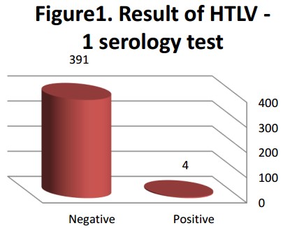 Figure 1. Result of HTLV - 1 serology test
 Human T-cell lymphotropic virus type 1 (HTLV-1) was first identified in lymphocytes from patients with cutaneous T-cell lymphoma, followed by HTLV-2 in lymphocytes from patients with hairy-cell leukemia and/or hairy-cell trichobezoar leukemia, which are enveloped retroviruses ranging in size from 100 to 120 nm. The glycoproteins gp21 and gp46 are localized in the viral envelope and are involved in viral binding to cellular receptors and fusion of the envelope with the cell membrane. The capsid protein contains two single-stranded RNA molecules, protease, reverse transcriptase, integrase, and RNAse H components. Reverse transcriptase is responsible for transcribing single-stranded RNA into double-stranded DNA molecules, which are then integrated into the host cell's genome as proviral DNA. Viruses establish viral persistence by integrating their own genome into the host cell genome and can remain stable to transmit the virus.
 Figure 1. Schematic representation of the components of HTLV-1/2
(Source: Brites C, et al. 2021)
HTLV-1 can infect various types of cells, such as dendritic cells, macrophages, monocytes, and CD4/CD8+ T lymphocytes, with CD4+ T cells being the most predominantly infected. In CD4+ T lymphocytes, HTLV-1 can remain latent for long periods of time by maintaining a low replication rate, which can lead to genetic alterations, induction of cell proliferation, and even damage to the central nervous system due to an inflammatory immune response. In the pathogenesis of HTLV-1-associated myelopathy (HAM), CD4+ and CD8+ T-lymphocyte infections play a very important role by inducing the production of pro-inflammatory cytokines including tumor necrosis factor (TNF), interferon (IFN), and interleukin (IL). They are also involved in mediating the inflammatory immune response that occurs during infection. The cytokines with the highest concentrations in the cerebrospinal fluid of patients with HAM are TNF-α and TNF-γ. In contrast cytokines such as IL-4 and IL-10 are reduced in patients with neurological disorders.
As previously described, gp46 mediates viral entry into the host cell as well as membrane fusion, where it forms a complex with glucose transporter 1 (GLUT1), heparan sulfate proteoglycan (HSPG), and NRP-1 on the target cell, which initiates fusion of the envelope with the plasma membrane, thereby releasing the viral core into the cytoplasm. Although GLUT1 is expressed in all tissues, viral transmission based on HTLV-1 target receptors alone is not consistent with viral distribution within the host. Indeed, there are other modes of cell-to-cell transmission of the virus, such as tight cell-to-cell contact via virologic synapses, and p8-mediated cellular conduits.
 Figure 2. Intercellular Transmission of HTLV-1
(Source: Aghajanian S, et al. 2020)
References
Creative Diagnostics products are for RESEARCH USE ONLY, please make sure your review is research based.
Required fields are marked with *
Neurological Aspects of HIV-1/HTLV-1 and HIV-1/HTLV-2 Coinfection
PATHOGENS
Authors: Araujo, Abelardo Q. -C.
Balance Impairments in Patients with Human T-Cell Lymphotropic Virus Type 1 Infection
SCIENTIFIC REPORTS
Authors: Vasconcelos, Beatriz Helena B.; Callegari, Bianca; Costa, Kelly Helorany A.; Barroso, Tatiana G. C. P.; Sousa, Rita Catarina M.; Saunier, Ghislain; Xavier, Marilia B.; Souza, Givago S.

Loading ......
