Upper motor neuron degeneration is typical of many neurodegenerative diseases, such as amyotrophic lateral sclerosis (ALS), hereditary spastic paraplegia (HSP), and primary lateral sclerosis (PLS). As one of the most complex nervous systems, the motor neuron system is mainly responsible for the initiation and modulation of voluntary movements. Among them, mouse cortical spinal motor neurons (CSMN) and human Betz cells are the key cortical components of this pathway. They can collect and integrate many different types of cortical input signals, convert them into information, and pass it to spinal cord targets to initiate voluntary movements. Previous studies have found that upper motor neuron degeneration is an early event in ALS, and that the cortical components of the disease show significant early signals before patients develop symptoms.

The mutation of the SOD1 gene is the most important discovery in the field of ALS. The establishment of a mouse model of SOD1 can be used to reveal the underlying mechanisms leading to the fragility and degeneration of motor neurons. Even if the lack of SOD1 does not cause degeneration, mutant SOD1 still exhibits toxic functions, such as causing oxidation and endoplasmic reticulum stress, axonal transport defects, and mitochondrial problems. Through further research on the SOD1 mutation model, more genes related to ALS have been discovered, among which Proflin 1 (PFN1) gene is one of them. Mutations in the PFN1 gene have been detected in ALS patients, and a Proflin mouse model has been generated to uncover the underlying mechanisms that cause motor neuron degeneration. In addition, mutations in the TARDBP gene were detected in ALS patients. The gene encodes a DNA / RNA binding protein, the TDP-43 protein. This protein is involved in many cellular functions, the most important of which is RNA metabolism. TDP-43 protein can regulate various aspects of RNA-related functions, including RNA processing, miRNA biogenesis and RNA splicing. TDP-43 pathology is defined as protein aggregation including phosphorylated forms of TDP-43 protein, and it is one of the most common protein pathological phenomena in the cortex of patients with ALS and ALS / FTLD. It is important to note that even in the absence of mutations in the TARDBP gene, TDP-43 pathological phenomena were observed in the brains of patients, including those in patients with mutations in their PFN1 gene. Under this pathological condition, the upper motor neurons of mice and humans also became fragile, and both CSMN and Betz cells showed nuclear membrane, ER, and mitochondrial defects.
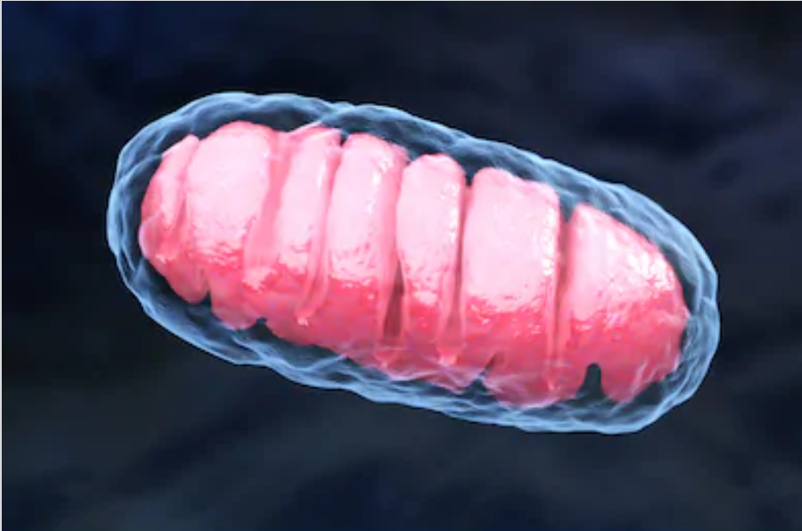
It is found that mitochondrial defect is one of the problems appearing in many neurodegenerative diseases through comparison of various neurodegenerative disease models. Previous studies have shown that mitochondria show ultrastructural defects in upper motor neurons in ALS patients and mouse models of ALS. In order to further study whether these mitochondrial defects are limited to the upper motor neurons in the motor cortex, whether the type and extent of mitochondrial defects are comparable among different underlying mechanisms, and when they start early. The researchers used neonatal mice at day 15 postpartum to assess any early mitochondrial damage that occurred in CSMN caused by TDP-43 pathology, mSOD1 toxicity, and lack of profilin function. Ultimately, researchers used immunocoupled electron microscopy to reveal the existence of a new type of self-destructive mitochondria. Because this pathway of mitochondrial degradation is different from previous mitochondrial damage, this phenomenon has been called “mitoautophagy” by researchers to emphasize that mitochondria can self-destruct without being engulfed by lysosomes or autophagosomes. Further research found that this phenomenon occurs very early in upper motor neurons, and mainly occurs in CSMN in mice. Therefore, mitochondrial abnormalities found during postnatal development may indeed be one of the first defects that lead to the fragility of upper motor neurons, especially in the context of TDP-43 pathology.
